What Does Skin Cancer Look Like – Early Signs and ABCDE Guide
Skin cancer is the most common form of cancer in many countries, yet recognizing its appearance remains challenging for many people. From pearly bumps to scaly patches, the visual signs of skin cancer vary widely depending on the type and stage. Understanding what to look for can make the difference between early treatment and more complex interventions later.
This guide provides a comprehensive overview of what skin cancer looks like across its main forms, including practical identification tools like the ABCDE rule. Whether you are checking a new spot on your arm or evaluating a changing mole on your face, the information here aims to help you understand when professional evaluation is warranted.
The three primary types of skin cancer—melanoma, basal cell carcinoma (BCC), and squamous cell carcinoma (SCC)—each have distinct visual characteristics, though they often share common warning signs. Learning to recognize these patterns empowers individuals to take prompt action when something suspicious appears on their skin.
What Does Skin Cancer Look Like?
Visual markers following the ABCDE criteria for melanoma and other characteristic lesions
Distinct appearances for melanoma versus basal cell versus squamous cell carcinomas
UV exposure history, skin type, and demographic influences on cancer presentation
Self-examination procedures and guidance for scheduling professional evaluations
Early Warning Signs
Skin cancer frequently develops in sun-exposed areas such as the face, neck, ears, scalp, and arms, though it can appear anywhere on the body. Early detection relies on recognizing changes that deviate from normal skin patterns. Non-healing sores that persist for weeks or longer often signal underlying skin damage that warrants medical attention.
Bleeding or oozing from a spot that seems minor, itching or pain in a localized area, and sudden appearance of new growths all merit closer inspection. The Skin Cancer Foundation emphasizes that persistent symptoms lasting more than a few weeks should prompt a consultation with a healthcare provider.
Common Appearances
While each type of skin cancer has specific features, common presentations include raised or flat growths, patches with altered texture or color, and sores that crust over without healing. Growths may appear pearlescent, scaly, crusty, or mole-like depending on the cancer type and the individual’s skin characteristics.
| Type | Typical Appearance | Prevalence | Early Survival Rate |
|---|---|---|---|
| Melanoma | Irregular mole with uneven coloring | Approximately 1% of skin cancers | 99% when detected early |
| Basal Cell Carcinoma | Pearly or waxy bump, often with visible blood vessels | About 80% of skin cancers | Nearly 100% with treatment |
| Squamous Cell Carcinoma | Red, scaly patch or firm nodule with crust | Around 20% of skin cancers | 95% or higher |
| Actinic Keratosis | Rough, sandpaper-like texture | Precancerous lesion | Manageable with early intervention |
| Invasive Melanoma | Evolving mole with multiple colors | Rarer but more aggressive | Depends on stage at detection |
According to the American Academy of Dermatology, one in five Americans will develop some form of skin cancer in their lifetime, making awareness and early recognition critically important health knowledge.
What Is the ABCDE Rule for Spotting Skin Cancer?
The ABCDE rule provides a simple framework for evaluating moles and growths for potential melanoma. This identification tool helps individuals distinguish between typical moles and those that may require professional assessment. The rule applies specifically to melanoma detection but offers useful general guidance for monitoring any skin changes.
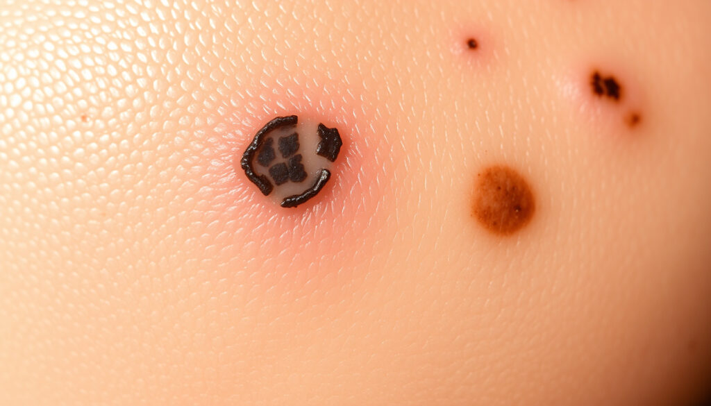
Asymmetry and Border Irregularity
A normal mole is typically symmetrical, meaning that if you draw a line through its center, both halves look similar. When one half of a mole or growth differs significantly from the other, asymmetry becomes a warning flag. Border irregularity complements this by describing edges that appear ragged, notched, blurred, or irregular rather than smooth and well-defined.
These characteristics often develop gradually, which is why the Mayo Clinic recommends performing regular self-examinations to establish a baseline for your specific skin markings and detect changes over time.
Color Variation and Diameter
Color variation within a single lesion suggests potential concern. While ordinary moles usually display a uniform single shade of brown, melanoma may contain multiple colors including black, brown, red, white, blue, or gray. The presence of two or more distinct colors within one growth warrants closer examination.
Diameter serves as another indicator. Most melanomas exceed six millimeters in diameter—roughly the size of a pencil eraser—though they can be smaller when first developing. The Cleveland Clinic notes that any growth larger than this threshold should be evaluated, particularly if other warning signs are present.
Evolving Changes
Evolution refers to any change in a mole or skin growth over time. This includes changes in size, shape, color, elevation, or surface texture. Symptoms such as itching, tenderness, bleeding, or oozing also qualify as concerning evolutions. A mole that looks different from your others or that is changing in any way merits professional attention.
Setting a monthly reminder to examine your skin helps establish familiarity with your unique pattern of moles and spots. The earlier changes are noticed, the earlier medical evaluation can occur.
How Do Different Types of Skin Cancer Look?
Each of the three main skin cancer types presents with distinctive visual features. Understanding these differences assists in identifying which type of cancer might be present, though definitive diagnosis requires professional medical evaluation and often laboratory testing.
Melanoma Characteristics
Melanoma is less common than other skin cancers but tends to be more aggressive when it develops. It frequently originates within an existing mole or appears as a new dark spot on the skin. The ABCDE rule applies most directly to melanoma identification, focusing on asymmetry, border, color, diameter, and evolving changes.
Stage 1 melanoma is typically thin—measuring less than one to two millimeters—and remains localized to its original site. When caught at this stage, cure rates are exceptionally high. Images of early melanoma often show asymmetric moles with multiple colors or irregular borders that distinguish them from benign growths.
Basal Cell Carcinoma Features
Basal cell carcinoma represents the most frequently diagnosed form of skin cancer. It typically develops as slow-growing lesions on skin that receives regular sun exposure. BCC often presents as a pearly, waxy, or flesh-colored bump with visible tiny blood vessels running through it.
Other BCC presentations include open sores that do not heal and may bleed, ooze, or crust over before seemingly healing and returning. Reddish patches of irritated skin, small pink growths with raised rolling edges, and scar-like areas with poorly defined borders all characterize different BCC subtypes.
Squamous Cell Carcinoma Signs
Squamous cell carcinoma often emerges from precancerous lesions called actinic keratoses, which appear as rough, scaly spots on sun-damaged skin. SCC itself typically manifests as firm red nodules or flat scaly patches with crusting. The lesions frequently develop a raised rim with a crusted center, sometimes resembling a volcano.
According to the Cancer Treatment Centers of America, SCC commonly appears on sun-exposed areas including the face, ears, neck, scalp, and hands. When SCC is identified early, treatment outcomes are excellent, with survival rates exceeding ninety-five percent.
What Does Skin Cancer Look Like on Different Skin Tones?
A common misconception holds that skin cancer primarily affects people with fair skin. Research published in dermatology journals confirms that skin cancer occurs across all skin tones, though presentations may differ significantly. Individuals with darker skin may experience diagnostic delays partly because cancerous lesions are often darker and may resemble common benign conditions.
On Darker Skin
On skin of color, basal cell carcinoma frequently appears pigmented rather than pearly or flesh-colored. Lesions may present as dark brown, black, blue, or multi-toned firm bumps that could be mistaken for moles, freckles, or age spots. This pigmentation pattern differs markedly from typical fair-skin presentations.
Squamous cell carcinoma on darker skin may also show atypical coloring and location patterns. The American Academy of Dermatology advises that photo galleries documenting skin cancer on diverse skin tones remain valuable resources for recognizing these variations.
Body Location Variations
Skin cancer on darker skin tones often develops in less sun-exposed areas such as the palms, soles, groin, and beneath fingernails or toenails. This distribution pattern differs from fair-skin presentations, which concentrate heavily on traditionally sun-exposed regions.
Any spot on the palms, soles, or mucous membranes that appears suspicious deserves prompt professional evaluation. These areas are less commonly monitored but represent known locations for skin cancer in individuals with darker complexions.
What Are the Stages and Unusual Presentations of Skin Cancer?
Skin cancer progresses through distinct stages when left untreated, though early identification typically occurs before advanced stages develop. Understanding stage progression and recognizing unusual presentations helps individuals appreciate the importance of timely medical consultation.
Early Stage Spots
Stage 1 skin cancers are thin and localized, often appearing as subtle pink, red, or brown bumps, small scaly patches, or evolving moles that catch attention through change rather than dramatic initial appearance. These early-stage lesions respond extremely well to treatment, with basal cell and squamous cell carcinomas achieving near-complete cure rates when addressed promptly.
Pimple-Like or Precancerous Lesions
Some skin cancers initially resemble pimples or other common benign conditions. Basal cell carcinoma may present as shiny nodules that look similar to acne pimples but persist and grow rather than resolving. Squamous cell carcinoma sometimes develops as firm red nodules with crusting that could be mistaken for irritated blemishes.
Growths that resemble pimples but continue to enlarge, bleed, or fail to heal after several weeks should be examined by a medical professional. Persistence and change distinguish potentially cancerous lesions from ordinary acne.
Precancerous actinic keratoses appear as rough, scaly spots that feel like sandpaper under the fingertip. These lesions represent direct precursors to squamous cell carcinoma in many cases. Dermatologists recommend treating actinic keratoses to prevent progression to invasive cancer.
How Skin Cancer Develops Over Time
Understanding the typical progression timeline of skin cancer reinforces the value of early detection and intervention. While individual experiences vary, general patterns help contextualize how precancerous changes develop into invasive disease.
- Ultraviolet exposure accumulates over years, causing cellular damage that may not become visible immediately. This cumulative sun damage forms the foundation for future cancer development.
- Precancerous lesions such as actinic keratoses may appear within months to years of significant sun exposure. These rough patches represent cellular changes that have not yet become cancerous.
- Visible cancer spots can develop within weeks to months once precancerous lesions progress to invasive cancer. The timeframe varies considerably based on individual factors.
- Without treatment, some skin cancers may eventually spread to surrounding tissues or more distant body areas. This metastatic potential underscores why early intervention matters.
What Is Established Versus Uncertain About Skin Cancer Identification?
Self-examination and careful observation provide valuable tools for identifying potential skin cancers, yet certain limitations exist that merit acknowledgment. Understanding what medical science has firmly established versus areas where uncertainty remains helps readers approach the information appropriately.
Skin cancer types have characteristic visual appearances; the ABCDE rule identifies melanoma features reliably; early detection significantly improves outcomes; UV exposure causes most skin cancers; regular self-examination supports early identification
Self-examination cannot replace professional diagnosis; biopsy confirms or rules out cancer definitively; some presentations defy typical categorization; AI-based detection tools show promise but require validation; individual risk factors influence disease patterns in ways that remain incompletely understood
Professional medical evaluation, including possible biopsy, provides the only definitive diagnosis. Self-monitoring serves as an important complement to regular dermatologist visits rather than a replacement for professional assessment.
The Importance of Context in Understanding Skin Cancer
Skin cancer does not develop in isolation. Understanding the risk factors and prevention context that surround skin cancer helps individuals appreciate why certain patterns of sun exposure and protective behaviors matter for long-term skin health.
Ultraviolet radiation from sunlight or tanning beds causes approximately ninety percent of skin cancers, according to the Skin Cancer Foundation. This strong connection between UV exposure and cancer development provides clear direction for prevention strategies. Fair skin, history of sunburns, excessive sun exposure, indoor tanning use, and genetic factors all influence individual risk levels.
Regular daily sunscreen application, wearing protective clothing, seeking shade during peak sun hours, and avoiding indoor tanning equipment represent evidence-based prevention approaches that reduce skin cancer risk over time.
Expert Sources and Professional Guidance
Medical organizations including the American Academy of Dermatology, the Skin Cancer Foundation, and major research institutions publish guidelines and educational materials on skin cancer identification and prevention. These organizations employ board-certified dermatologists and researchers who contribute to advancing understanding of skin cancer presentation and treatment.
“The ABCDE rule serves as a critical tool for identifying melanoma early, when treatment is most effective. Anyone who notices any of these warning signs should make an appointment with a board-certified dermatologist promptly.”
— American Academy of Dermatology public guidance on skin cancer detection
Healthcare providers with dermatology training offer expertise in recognizing subtle presentations and determining when biopsy or further testing is appropriate. Annual skin examinations by qualified professionals complement personal monitoring efforts.
Key Takeaways for Monitoring Your Skin
Recognizing what skin cancer looks like across its various forms empowers individuals to monitor their skin effectively and seek appropriate care when concerning signs appear. The visual characteristics covered in this guide provide a foundation for informed self-examination.
Monthly skin self-checks, prompt attention to changing or unusual spots, and regular professional dermatology visits form a comprehensive approach to skin cancer early detection. When in doubt, scheduling a professional evaluation provides peace of mind and ensures that any necessary treatment occurs at the earliest possible stage.
Frequently Asked Questions
When should I see a doctor for a skin spot?
Consult a healthcare provider for any new growth, sore that does not heal within three to four weeks, spot that itches or bleeds, or any mole that changes in size, shape, or color. The CDC recommends professional evaluation whenever skin changes seem unusual or persistent.
What does precancerous skin look like?
Precancerous actinic keratoses appear as small rough, scaly patches that feel like sandpaper. They often develop on sun-exposed skin and may be easier to feel than see. These lesions can progress to squamous cell carcinoma if left untreated.
Can skin cancer look like a pimple?
Yes. Some basal cell and squamous cell carcinomas initially resemble persistent pimples. The key distinction involves persistence and change—if a pimple-like growth does not resolve within a few weeks, grows larger, or bleeds easily, professional evaluation is advisable.
What does stage 1 skin cancer look like?
Stage 1 skin cancers appear as subtle bumps, small patches, or evolving moles that are typically less than one to two millimeters thick and remain localized. These early-stage lesions often look similar to benign growths, which is why change over time serves as an important warning signal.
How does skin cancer start?
Skin cancer typically begins when ultraviolet radiation damages skin cells, causing genetic mutations that allow abnormal cells to proliferate. These damaged cells may first appear as precancerous lesions or subtle changes that gradually develop into visible cancers over months to years.
What does skin cancer look like on black skin?
On darker skin tones, skin cancer often appears as dark brown or black firm bumps, scaly patches, or non-healing sores that may resemble moles or age spots. Cancers on people of color frequently occur in less sun-exposed areas such as palms, soles, and groin region.
Does skin cancer always come from moles?
Not always. While melanoma commonly develops within existing moles, basal cell and squamous cell carcinomas typically arise from normal skin or precancerous lesions rather than moles. New spots that appear in adulthood warrant closer attention regardless of their appearance.
What are the signs of skin cancer on the arm?
Skin cancer on the arms commonly appears as firm red nodules, scaly patches, open sores that do not heal, or pearly bumps with visible blood vessels. The arms receive significant sun exposure, making them frequent sites for all three main skin cancer types.
More related posts
 Yokohama FM vs Liverpool: Final Score, Goals & Highlights
 Clint Rice Net Worth: MAFS Golf Star’s Fortune Explored
 John Lennon: Death, Last Words & $31,000 Tooth Facts
 Tuna Pasta Recipe: 5 Easy Variations for a Quick Dinner
 Online Services for Agents – Top 2025 CRM and Lead Tools
 Geelong Adventure Park – Essential 2025 Visitor Guide
 Water Cycle Diagram: 5-7 Steps Explained (Simple to Advanced)
 Pep Guardiola 2025: Wife Reunion & Man City Future